X-RAY FREQUENCY AND SAFETY
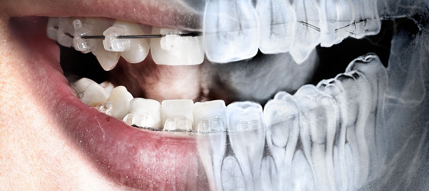
In dental procedures, sometimes requires jaw and teeth’s x-ray views. However it is safe or harmfull to our body. First let's look at what “x-rays” are and why they're used. X-rays are a form of electromagnetic radiation, just like light, except they just have a much shorter wave-length. X-rays are a form of “ionizing” radiation which basically means they can penetrate body tissues which is what generally prompts concern. However it is just this property which makes them important diagnostic tools. They can penetrate soft tissues like skin and gums much more readily than hard tissues like bone and teeth causing different degrees of shadows. The shadows can be captured on film or digital receivers and are called radiographs (x-ray pictures). Because today's x-ray machines and image capturing techniques are so sensitive, the amount of radiation needed for diagnosis is negligible, almost next to nothing compared to what you get from every day background radiation.

First a little science: A millisievert (mSv), named after Dr. Rolf Sievert, famous for studying the biological effects of radiation, is the unit of measurement that allows for comparison of doses from different x-ray sources. We use this measurement to help determine what we call the effective dose (E), a way of calculating the safety factor of each x-ray exposure. Since we know our annual background exposure to natural x-radiation (all around us) is from 2 to 4.5 mSv, and more if you like to take airplane rides, we can then make a comparison to dental x-ray examinations.
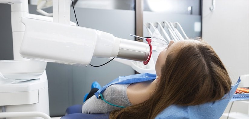
Dental radiographs are completely safe; the average single digital periapical film (peri-around, apical-root end of a tooth) is equal to 1 microsievert (µSv) i.e. one thousandth of an mSv. For four bitewing radiographs, traditionally used to image the back teeth for decay (the little tabs you bite on are called bite-wings), the exposure is 4 µSv. The x-ray machines take images of only the necessary structures, so there is no scatter of the x-rays to other tissues. Your dentist may even take the precaution of making you wear a lead apron to shield the rest of your body.
A full mouth series of 18-20 radiographs (all the teeth) using “D” speed film is equal to about 85 µSv. “D” speed film, long considered the gold standard in dental imaging, exposes the teeth to the “highest” radiation dose. Even with this, radiographs taken using “D” speed film equal just seven to ten day's background radiation. Today, most dentists taking a full series use “F” speed film or digital receivers. These receptors now equal the quality of “D” speed film and are much more sensitive to x-rays thus reducing exposure by as much as 80%. This means a full mouth series of radiographs taken with “F” speed film or digital receivers equal just a half day's background radiation.

How often radiographs are needed depends on your individual health needs. Your dentist will review your history, examine your mouth and teeth then decide whether you need radiographs and what type. For new patients, an overall screening is typically indicated using a very low x-ray dosage panoramic radiograph. As the name implies this gives a panoramic screening view of the head, neck, sinuses, jaw bones, teeth and more. It allows a determination of overall health status of all these structures and is used to detect hidden and importantly, “silent” conditions that don't cause symptoms (at least not until late stages) like cysts, cancers and of course tooth decay and gum disease.

After overall oral health and dental status have been preliminarily determined, more detailed images can be taken using smaller radiographs of individual teeth called periapicals, or bitewing radiographs for example. These routine pictures taken with a standard technique give your dentist a multitude of information about tooth decay and periodontal (gum) disease (which results in bone loss), all imaged in great detail with extraordinarily little radiation.
Once your dentist has a track record with you and has made an evaluation of your individual risk for cavities or gum disease, he/she will be able to assess the interval and type of radiographs necessary to monitor you over time. At your age, it would not be uncommon for a dentist to recommend bitewing radiographs on an annual basis, but this will depend largely on your history, decay experience, number of fillings and many other factors which I am not party to.
You should discuss your concerns directly with your dentist and ask him/her to review them with you, you'll learn a great deal and probably feel a lot more comfortable when you have direct answers to your questions.
Endişelerinizi doğrudan diş hekiminiz ile görüşmelisiniz, bu sorulara doğrudan cevap verildiğinde kendinizi daha iyi hissedeceksiniz.